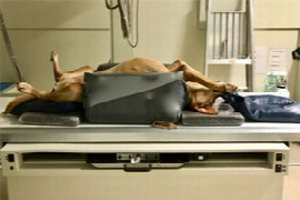
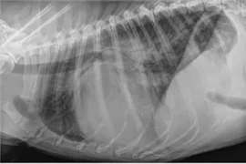
Radiographic positioning is essential for correct identification and diagnoses of lesions on radiographs. It is essential to understand how to acquire correctly positioned orthogonal radiographs and how positioning results in the projected image. The webinar will focus on how to position dogs and cats for the optimal radiograph of the thorax, abdomen and selected musculoskeletal anatomy. The webinar will expand on how to position these patients to minimise x-ray exposure to both the staff and patient, what to look for in a correctly positioned radiograph, tips on how to problem solve incorrectly positioned radiographs, and will introduce less new projections that may offer further diagnostic value to radiographs.
Recording from 29 January 2019
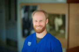
Rob is a Massey University trained veterinarian from Zimbabwe. He completed his ANZCVS Memberships in Small Animal Radiology in 2015 and completed his residency training in veterinary diagnostic imaging at U-Vet Werribee Animal Hospital this year (2018). He currently works as a diagnostic imaging registrar at U-Vet as he prepares to write his fellowship exam in 2020. His current research projects include image characteristics of the pancreas and body composition determination by various imaging modalities. He is also very enthusiastic about vascular anomalies and neurologic imaging. Preceding his residency, he worked in general practice in Adelaide, Cairns and for a short period, Jaipur, India. He enjoys combining travel with work, having travelled to India twice and to China to assist in small animal welfare clinics, camel, and horse welfare education, and to volunteer at the Animal Asia Centre in Chengdu. When not at work, Rob enjoys Crossfit and running with his always energetic German Short-haired Pointer