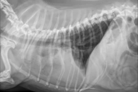
Thoracic radiographs are commonly performed in small animal practice, but interpreting lung changes can still be challenging. This webinar will help general practitioners recognise the main canine and feline pulmonary patterns, including bronchial, interstitial, alveolar and vascular changes utilising real life cases to assist learning. Using real clinical cases and interactive image interpretation questions, participants will learn how lesion distribution, associated thoracic findings and clinical history can help prioritise differential diagnoses. The aim is to build a structured, practical approach that can be applied immediately to interpreting lung changes in coughing, dyspnoeic and systemically unwell patients in everyday practice.
Language: English
Accredited by the Austrian Chamber of Austrian Chamber of Veterinarians (ÖTK) for:
- 2.00 hours – General Education
- 2.00 hours – Specialist Veterinarian (FTA) Small Animal Internal Medicine
- 2.00 hours – Specialist Veterinarian (FTA) in Small Animals
Accredited by ATF
- 1 hour according to § 10 of ATF-Statutes
The continuing education (CE) hours are accredited, provided that the knowledge assessment is successfully completed within the specified period. The accreditation is valid from 23 September 2026 to 22 September 2027.
Referent:In

Pete Mantis
Panagiotis “Pete” Mantis DVM, PhD, DipECVDI, FHEA, FRCVS
Pete joined Dick White Referrals in 2017 and was appointed Head of Diagnostic Imaging in 2019. He is a European and RCVS Specialist in Veterinary Diagnostic Imaging, a Fellow of the Higher Education Academy, and a Fellow of the Royal College of Veterinary Surgeons.
He is the founder of Vet-Learn.com and is a regular author, speaker and CPD tutor in small animal radiology, ultrasonography, computed tomography and magnetic resonance imaging. Pete has a particular interest in making diagnostic imaging practical, clinically relevant and accessible for vets working in everyday small animal practice.