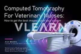
This webinar is tailored to Veterinary Nurses who have a keen interest in diagnostic imaging, and are utilising Computed Tomography in practice. The 45 minute presentation will provide an overview of how a CT scanner works, as well as guidance and recommendations for positioning and acquisition techniques, giving attendees a foundation in the use of this advanced imaging modality. There will the opportunity for attendees to ask questions relating to the use of Computed Tomography in Veterinary Practice.
Language: English
Recording from December 10, 2024
1 hour according to § 10(2) of the ATF Statutes as mandatory continuing education for ATF members.
ATF hours are recognized by the ÖTK as educational hours.
Recognition according to the guidelines of the Society of Swiss Veterinarians is possible.
The accreditation of CE Points is approved under the condition of a positive completion of the test in the specified period and is valid from December 10, 2024 to December 10, 2025.
Speaker:
Ashley Moors
Ashley Moors, FdScVN GradDipVN PGCertVetEd FHEA RVN, Veterinary Radiographer, Royal Veterinary College
Ash graduated from the Royal Veterinary College, UK with an FdSc in Veterinary Nursing in 2010, and worked in First Opinion Practice in Dorset with access to Low Field MRI and Digital Radiography Systems.
He returned to the RVC in 2012, undertaking the Graduate Diploma in Clinical and Professional Veterinary Nursing whilst continuing to work in First Opinion Practice, choosing to undertake the surgical nursing and diagnostic imaging elective modules, which further developed his interest in radiography. During this period he built and coded a custom image storage system for the practice he worked at, allowing query and retrieval of digital imaging studies.
Ash joined the RVC's Queen Mother Hospital for Animals in 2016 as a Veterinary Radiographer, working exclusively in the Radiology department and performing a range of diagnostic imaging studies, including Fluoroscopy, CT and MRI. He is currently Administrator for the RVC's Vendor Neutral Archive storage system, overseeing a system of interconnected storage and viewing platforms that serve all clinical imaging equipment across both RVC campuses at Hawkshead and Camden.
He has been involved in teaching Veterinary Nurses at post graduate level, as well as providing CPD courses focused on Diagnostic Imaging, and has a particular interest in the provision of practical radiography skills to both Veterinary Nurses and Veterinary Surgeons, as well as the use of CT and MRI data for 3D models to assist with student education and owner understanding.