Webinars & e-learning courses All webinars

In house cytology of skin masses- tips and tricks

In house cytology of skin masses- tips and tricks

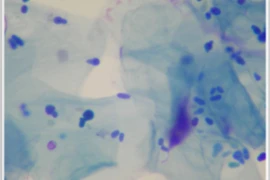
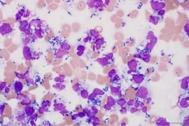
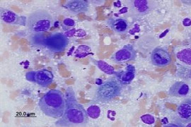
Have you been frustrated by your cytology samples and getting the right cytologic diagnosis? Here is your opportunity to develop those skills. This webinar will outline the tips and tricks to getting a cytologic diagnosis. We will start with how to increase your chances of a diagnostic quality sample – covering sample collection and preparation. We follow with an outline of a systematic approach to evaluation of cytologic specimens – identifying inflammation versus neoplasia and criteria of malignancy, and categorization of neoplasia as epithelial, mesenchymal, round cell, and neuroendocrine. We will then explore some of the exceptions to our cytology rules and guidelines of when to seek out help from an expert. All complemented with lots of pictures of clinical cytology specimens.

Recording from 21.02.2019

Speaker:
Nat.jpg

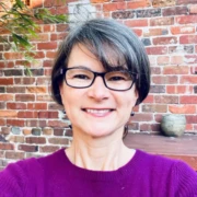
Natalie Courtman

DVM, BVSc, MANZCVS, MVS, GCUT, DipACVP
Natalie graduated from the University of Sydney and spent nine years in small animal practices in Australia and the UK before embarking on her pathology career at the Royal Veterinary College in the UK. She is a Diplomate of the American College of Veterinary Pathologists in clinical pathology, and has a Masters in Veterinary Clinical Studies from Murdoch University and membership of the ANZCVS in Small Animal Medicine and Pathobiology. She has worked in private veterinary laboratories and at the University of Melbourne training DVM students and residents. Her passion is helping vets and vet nurses find and understand the clues that will help them diagnose disease in their patients. 

Booking information

Duration: 0:51 h
Speaker: Natalie Courtman
from 1
54.90 US$
(inkl. tax)

You might also be interested in

Crimson crisis 300x200 (1).jpg
Craig Breheny
1:01 h

Crimson Crisis - Decoding Immune-Mediated Haemolytic Anaemia

Diagnosing IMHA, ruling out triggers, and then deciding on the best treatment strategy can be a challenge. In this talk Craig Breheny will take you from presentation to recheck with some clinical tips.


Image 22.03 300x200.jpg
Astrid Oscos Snowball
0:53 h

Pathology - Microscope Technique - Cell Morphology and Interpretation for Nurses

The correct use of the microscope is important to optimize microscopic visualisation of cytology specimens. Cell identification is useful in diagnosing and ruling out certain conditions, narrowing the list of differential diagnoses.
3476 300x200 (1).png
01.12.2026 (UTC)
09:00 - 10:00 (UTC)

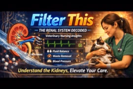
Filter This - The Renal System Decoded for Vet Nurses

A clear, practical understanding of kidney anatomy and physiology revealing how these vital organs filter, balance, and protect the body which are essential for everyday patient care with Melissa Shoard.