Webinars & e-learning courses All webinars

Canine Epiphyseal Disease

Canine Epiphyseal Disease

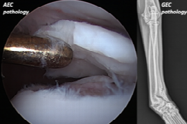
The epiphysis is located at either end of long bones and is evident within the fetus approximately four weeks post conception. During growth, the epiphysis is comprised of specialized cartilage, including articular epiphyseal cartilage (AEC) and growth plate epiphyseal cartilage (GEC), together accounting for longitudinal growth of long bones. In maturity, most of the epiphyseal cartilage is replaced with trabecular bone, encased in a thin shell of cortical bone with hyaline cartilage persisting at the joint surface.

Epiphyseal pathology sustained during growth can have profound lifelong consequence. This webinar reviews common epiphyseal pathologies, including articular and growth plate osteochondrosis and epiphyseal trauma. A focus on diagnosis, treatment and expected outcomes with practical tips and tricks for use in your practice is presented.

Recording from 28.05.2020

Speaker:

Webp.net-resizeimage__2_.jpg

Ricky Cashmore

DVM, BVSc, FANZCVS (SAS)

Following graduation from the University of Melbourne (2005) Ricky worked in general practice for 18 months prior to completion of a rotating internship at the University of Cambridge, surgical internship at the Veterinary Specialist Centre and surgical residency at the Small Animal Specialist Hospital in Sydney (2010-2013). Ricky remained at SASH until 2017 before joining the Southpaws Speciality Surgery for Animals team in Melbourne (current practice).
Ricky is a Fellow of the Australian New Zealand College of Veterinary Scientist in Small Animal Surgery. He has published and presented around the world with a strong and active interest in clinical and biomechanical research relating to the canine hip, stifle and spine, the focus of his current and previous research projects. He is a past recipient of the prestigious VOS Wade O Brinker Orthopaedic Resident Research Award (presented WVOC, Colorado 2014) and previous examiner for the ANZCVS small animal surgical chapter. He thoroughly enjoys all aspects of teaching, having taught at numerous workshops and conferences nationally and internationally while mentoring many residents and interns over the years.
Ricky’s primary surgical interests lie in orthopaedics and include arthroscopy, partial and total joint replacement, minimally invasive fracture repair and neurosurgery.
Booking information

Duration: 1:05 h
Speaker: Ricky Cashmore
from 1
57.60 US$
(inkl. tax)

You might also be interested in

300x200.jpg
Elsa Beltran
1:08 h

The clinical approach to paraplegia in dogs

Myelopathies resulting in paraparesis/paraplegia in dogs and cats are commonly encountered in clinical practice.  This lecture reviews the clinical and diagnostic approach the dogs presented with paraparesis/paraplegia in describes pathophysiology, clinical presentation, diagnosis, treatment, outcome and prognosis of specific disorders.
Webp.net-resizeimage (7).jpg
Dorothee Krainer
0:55 h

Lumbosacral Stenosis

This vet-webinar reviews the aetiology, diagnosis and management of degenerative lumbosacral stenosis. Lumbosacral stenosis occurs when soft tissue and/or bony changes impinge on the nerve roots or vasculature of the cauda equina. The impingement can be worsened in conjunction with abnormal motion of the lumbosacral joint. Lumbosacral stenosis occurs most frequently in middle-aged dogs of medium to large breed, and  German Shepherd dogs are predisposed to this disease. Common signs are lumbosacral pain, lameness, pelvic limb weakness and ataxia, and urinary incontinence. Diagnosis is based on clinical features and imaging studies. Depending on the severity of the compression but about 50% of dogs respond well to conservative treatments. Decompressive surgery is effective in most patients.
decision-making-for-hip-dysplasia-what-to-advise-and-when.jpeg
Arthur House
0:40 h

Decision making for hip dysplasia – what to advise and when

Several options are available for the management of hip dysplasia. To accurately advise dog owners with a pet that suffers from hip dysplasia, the mechanism of action and the expected outcomes for each treatment option need to be understood. Many of the treatment options available for hip dysplasia have very specific applications and hence can not be used in all cases. This lecture will outline the options available and there application so that a treatment algorithm that can be created for each pet.