Webinars & e-learning courses All webinars

Lumbosacral Stenosis

Lumbosacral Stenosis

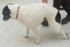
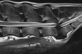
This vet-webinar reviews the aetiology, diagnosis and management of degenerative lumbosacral stenosis. Lumbosacral stenosis occurs when soft tissue and/or bony changes impinge on the nerve roots or vasculature of the cauda equina. The impingement can be worsened in conjunction with abnormal motion of the lumbosacral joint. Lumbosacral stenosis occurs most frequently in middle-aged dogs of medium to large breed, and  German Shepherd dogs are predisposed to this disease. Common signs are lumbosacral pain, lameness, pelvic limb weakness and ataxia, and urinary incontinence. Diagnosis is based on clinical features and imaging studies. Depending on the severity of the compression but about 50% of dogs respond well to conservative treatments. Decompressive surgery is effective in most patients.

Recording from 22 September 2020

Speaker:

Dorothee Image.jpg

Dorothee Krainer

DVM, Dipl.ECVS, European Specialist in Small Animal Surgery
Dorothee is an ECVS- and RCVS-recognised specialist in Small Animal Surgery. She enjoys all aspects of Small Animal Surgery including minimal invasive procedures and neurosurgery. Dorothee studied in Sydney, Budapest and Leipzig. Her doctorate was on BoneWelding® in spinal surgery, which she received from Zurich University. Dorothee trained her surgical skills in London, at UCD Dublin and at the VetMedUni Vienna, where she also worked several years on the orthopaedic service before she got appointed head of surgery at a leading referral hospital in Austria. Dorothee is a recognized international speaker and reviewer for several surgical journals. Her main research topic is brachycephalic syndrome. She currently works on her thesis for her professional master of business administration.

Booking information

Duration: 0:55 h
Speaker: Dorothee Krainer
from 1
57.60 US$
(inkl. tax)

You might also be interested in

golden-retriever_300x200.jpg
Hannah Capon, Anne Strattner, Larissa Postrak
1:23 h

Game Changers in Osteoarthritis Management

Sie betreiben bei Hunden mit OA multimodales Schmerzmanagement? Super! Aber vielleicht geht noch mehr. Wir sprechen über das häusliche Umfeld, Gelenkschutz und Hilfsmittel.
decision-making-for-hip-dysplasia-what-to-advise-and-when.jpeg
Arthur House
0:40 h

Decision making for hip dysplasia – what to advise and when

Several options are available for the management of hip dysplasia. To accurately advise dog owners with a pet that suffers from hip dysplasia, the mechanism of action and the expected outcomes for each treatment option need to be understood. Many of the treatment options available for hip dysplasia have very specific applications and hence can not be used in all cases. This lecture will outline the options available and there application so that a treatment algorithm that can be created for each pet.
canine-epiphyseal-disease.png
Ricky Cashmore
1:05 h

Canine Epiphyseal Disease

The epiphysis is located at either end of long bones and is evident within the fetus approximately four weeks post conception. During growth, the epiphysis is comprised of specialized cartilage, including articular epiphyseal cartilage (AEC) and growth plate epiphyseal cartilage (GEC), together accounting for longitudinal growth of long bones. In maturity, most of the epiphyseal cartilage is replaced with trabecular bone, encased in a thin shell of cortical bone with hyaline cartilage persisting at the joint surface. Epiphyseal pathology sustained during growth can have profound lifelong consequence. This webinar reviews common epiphyseal pathologies, including articular and growth plate osteochondrosis and epiphyseal trauma. A focus on diagnosis, treatment and expected outcomes with practical tips and tricks for use in your practice is presented.