Webinars & e-learning courses Intermediate All webinars

Reading thoracic radiographs of the dog and cat: how to go about?

<span>Reading thoracic radiographs of the dog and cat: how to go about?</span>

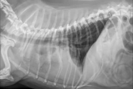
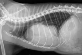
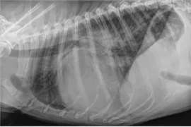
Thoracic radiography is one of the most common imaging examinations in general practice, yet many clinicians find the images hard to interpret with confidence. This one-hour webinar gives a practical, structured approach to reading canine and feline thoracic radiographs.
The session discusses the reading technique. It covers how to work through every image so that important findings are not missed. It then reviews the normal radiographic anatomy of the thorax in dogs and cats: the thoracic wall, pleural space, mediastinum, heart and great vessels, pulmonary vasculature and lungs. The webinar also presents simple rules of thumb to help decide whether an organ is normal. 
Attendees will leave with a systematic method to evaluate thoracic radiographs of dogs and cats and tips of how to evaluate normality that they can apply to their next thoracic study.

Language: English

An application for CE Hours is submitted. 

Speaker:

E_26_07_21_Radiology of the canine and feline lung made practical_Pete_200x200.png

Pete Mantis

Panagiotis “Pete” Mantis DVM, PhD, DipECVDI, FHEA, FRCVS

Panagiotis “Pete” Mantis graduated from the Faculty of Veterinary Medicine, Aristotle University of Thessaloniki, Greece, in 1994. He completed a residency in Veterinary Diagnostic Imaging at the Royal Veterinary College, London, and subsequently worked in both first-opinion and referral practices in London. After spending a year at the Swedish University of Agricultural Sciences in Uppsala, he returned to the Royal Veterinary College in 2000, where he became Senior Lecturer in Radiology.
Pete joined Dick White Referrals in 2017 and was appointed Head of Diagnostic Imaging in 2019. He is a European and RCVS Specialist in Veterinary Diagnostic Imaging, a Fellow of the Higher Education Academy, and a Fellow of the Royal College of Veterinary Surgeons.
He is the founder of Vet-Learn.com and is a regular author, speaker and CPD tutor in small animal radiology, ultrasonography, computed tomography and magnetic resonance imaging. Pete has a particular interest in making diagnostic imaging practical, clinically relevant and accessible for vets working in everyday small animal practice.
Booking information

Date 13.01.2027 (UTC)
Time 19:00 - 20:00 (UTC)
Speaker: Pete Mantis
from 1
57.60 US$
(inkl. tax)

You might also be interested in

Figure-4-web 300x200.png
Lily Hung
1:09 h

Telltale signs of cardiomegaly and heart failure on radiographs and TFAST

Lily Hung presents a review and updates on the current literature with top 5 tips to identify cardiomegaly on thoracic radiography. 
E_2026_02_19_Immune-mediated Polyarthritis - When to suspect, what to rule out and what to treat with_300x200-2.png
Craig Breheny
1:00 h

Immune-mediated polyarthritis - when to suspect, what to rule out and what to treat with

Immune-mediated polyarthritis can affect many body systems and prove a diagnostic challenge. With abundant underlying triggers, Craig Breheny will talk about how deciding on a work up plan, and the associated therapeutic interventions can be a challenge.
E_26_07_21_Radiology of the canine and feline lung made practical_300x200.png
Pete Mantis
1:09 h

Radiology of the canine and feline lung made practical

In this webinar with Pete Mantis you will learn to read thoracic radiographs.