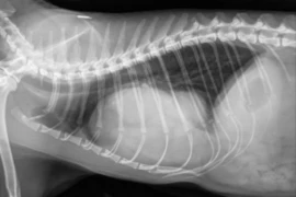
Thoracic radiography is one of the most common imaging examinations in general practice, yet many clinicians find the images hard to interpret with confidence. This one-hour webinar gives a practical, structured approach to reading canine and feline thoracic radiographs.
The session discusses the reading technique. It covers how to work through every image so that important findings are not missed. It then reviews the normal radiographic anatomy of the thorax in dogs and cats: the thoracic wall, pleural space, mediastinum, heart and great vessels, pulmonary vasculature and lungs. The webinar also presents simple rules of thumb to help decide whether an organ is normal.
Attendees will leave with a systematic method to evaluate thoracic radiographs of dogs and cats and tips of how to evaluate normality that they can apply to their next thoracic study.
Language: English
An application for CE Hours is submitted.
Speaker:

Pete Mantis
Panagiotis “Pete” Mantis DVM, PhD, DipECVDI, FHEA, FRCVS
Pete joined Dick White Referrals in 2017 and was appointed Head of Diagnostic Imaging in 2019. He is a European and RCVS Specialist in Veterinary Diagnostic Imaging, a Fellow of the Higher Education Academy, and a Fellow of the Royal College of Veterinary Surgeons.
He is the founder of Vet-Learn.com and is a regular author, speaker and CPD tutor in small animal radiology, ultrasonography, computed tomography and magnetic resonance imaging. Pete has a particular interest in making diagnostic imaging practical, clinically relevant and accessible for vets working in everyday small animal practice.