Webinars & e-learning courses All webinars

Reading thoracic radiographs of the dog and cat

Reading thoracic radiographs of the dog and cat

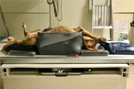
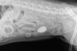
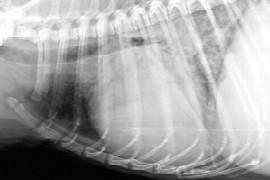
In this webinar we shall concentrate on the technique of reading thoracic radiographs of the dog and cat. Case examples will be discussed to provide a clear understanding of the reading technique and how to go about when you read thoracic radiographs.

Language: English

In case you have missed this webinar, you have the opportunity to watch a recorded version here.

This webinar qualifies for one (1) hour of continuing education.


Speaker:

Pete_small.jpeg

Pete Mantis

DVM, DipECVDI, FHEA, MRCVS

Pete is a European Specialist in Veterinary Diagnostic Imaging and an RCVS Recognized Specialist in Diagnostic Imaging. He is a regular author, speaker and tutor on the subjects of small animal radiology, ultrasonography, computed tomography and magnetic resonance imaging.
Pete is Senior Lecturer in Radiology at the Royal Veterinary College, University of London.

Booking information

Duration: 1:03 h
Speaker: Pete Mantis
from 1
54.90 US$
(inkl. tax)

You might also be interested in

radiographic-positioning-for-veterinary-nurses.jpeg
Robert Turner
0:57 h

Radiographic Positioning for Veterinary Nurses

Radiographic positioning is essential for correct identification and diagnoses of lesions on radiographs. It is essential to understand how to acquire correctly positioned orthogonal radiographs and how positioning results in the projected image. The webinar will focus on how to position dogs and cats for the optimal radiograph of the thorax, abdomen and selected musculoskeletal anatomy. The webinar will expand on how to position these patients to minimise x-ray exposure to both the staff and patient, what to look for in a correctly positioned radiograph, tips on how to problem solve incorrectly positioned radiographs, and will introduce less new projections that may offer further diagnostic value to radiographs.
E_26_03_10_ Humeral Condylar Fractures_300x200.png
Ian Faux
1:03 h

Humeral Condylar Fractures

Humeral condylar fractures are unique and challenging fractures. This lecture by Ian Faux will provide an overview with an emphasis on evidence based decision making and practical tips.
300x200.jpg
Catheryn Walsh
0:43 h

Diagnosing a Foreign Body

A discussion that shows the vital radiographic signs to look out for when deciding if a foreign body is present and whether to cut or not to cut. This will include real world case studies to help reinforce the teaching points.