

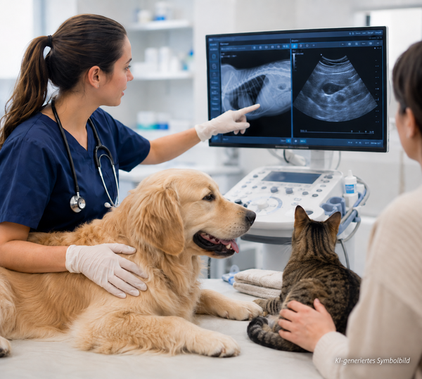
Diagnostic Imaging in Veterinary Practice
Diagnostic imaging shows internal structures but does not produce a diagnosis in isolation. History, examination and laboratory findings guide selection and interpretation.
The best examination answers the clinical question reliably and changes patient management; it is not automatically the most advanced technique.
Veterinary Imaging Courses at vet-webinar
vet-webinar offers live webinars, recordings, webinar packages and E-Learning on abdominal ultrasound, interpretation, artefacts and cross-sectional anatomy.
Learning That Supports Clinical Practice
Case studies presented by expert instructors show how imaging findings guide clinical decisions. Courses help veterinarians at every career stage expand their knowledge, skills and diagnostic confidence, including interns and residents working with small animals or horses.
Learn at Your Own Pace
Foundation, advanced and expert-level courses support professional growth and can be completed at your own pace. Dates, access details and included quizzes are listed on each course page.
RATO - Rationale and early clinical results
GI Ultrasound in dogs & cats
Ultrasound modular course
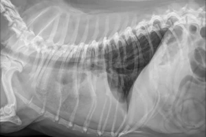
Practical lung radiology
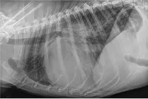
Cardiomegaly & Heart failure
Maximise Your Ultrasound!
Continuing Education That Makes a Difference in Clinical Practice
A technically good image is not yet a diagnosis.
What matters is knowing how reliable a finding is, which differential diagnoses to consider, and what the results mean for the next steps in your patient's diagnosis or treatment.
That's why our courses combine imaging techniques, practical skills, interpretation and clinical decision-making. Rather than simply showing abnormal images, they explain step by step how findings are obtained and how to interpret them in the context of the whole clinical case.
More than 90% of our team are veterinarians. We work closely with veterinary practitioners and speakers from around the world. Our goal is not just to provide scientifically sound continuing education, but to offer knowledge you can actually use with your next patient – helping you feel more confident in your clinical decisions and rediscover the satisfaction of practising good veterinary medicine.

Deepen Your Imaging Knowledge – and Explore Other Veterinary Specialties
Some questions can be answered with a single course. Other topics require practice, different clinical cases, and time to develop a deeper understanding.
With the vet-webinar Gold Subscription, you get access to webinars, webinar packages, and e-learning courses across our full range of included continuing education. This covers not only radiology and diagnostic imaging, but also internal medicine, cardiology, surgery, neurology, emergency medicine, and many other veterinary specialties.
Frequently asked questions
Diagnostic Imaging in veterinary practise

Diagnostic imaging shows internal structures but does not produce a diagnosis in isolation.
History, examination and laboratory findings guide selection and interpretation. Radiography, ultrasound, CT and MRI each have their strengths, depending on the patient and the clinical question.
The best examination answers the clinical question reliably and changes patient management: it is not automatically the most advanced technique.
Understanding what each imaging method can and cannot tell us is essential for making informed clinical decisions. It helps veterinarians choose the right examination, interpret findings in context and decide what to do next.
Which Diagnostic Imaging Modality Answers Which Question?
Ultrasound for Small Animals
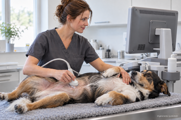
An ultrasound system alone does not guarantee a useful examination. Patient handling, systematic coverage and documentation determine reproducibility.
Abdominal Ultrasound in Dogs
In dogs, abdominal ultrasound supports gastrointestinal and urinary investigations, free fluid and masses. Changes require multiplanar assessment and clinical correlation.
Abdominal Ultrasound in Cats
In cats, small changes may be relevant and smaller organs require high frequencies. Gastrointestinal layering, pancreas, hepatobiliary system, kidneys and lymph nodes should be assessed together.
Veterinary Radiology: From Exposure to Findings
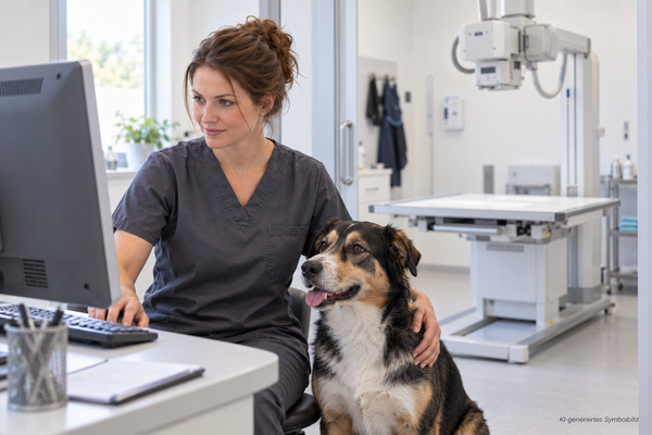
The diagnostic value of an X-ray begins before interpretation. Positioning, centring, exposure and motion determine whether structures are assessable.
Good radiographic technique is essential for obtaining images that allow reliable interpretation. Even small positioning errors or inadequate exposure can hide important details or create misleading findings.
Assess Radiographs Systematically
A fixed review sequence reduces missed findings. Describe location, size, shape, margin, opacity, distribution and relationships before developing differential diagnoses.
Reviewing all visible anatomical structures, rather than focusing only on the most obvious abnormality, helps identify subtle changes and incidental findings. Combining radiographic findings with the patient's clinical history and examination results is essential for reaching a meaningful diagnosis.
CT and MRI in Veterinary Medicine
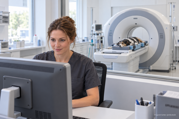
Advanced imaging often occurs in specialty hospitals. For referred patients, the practitioner remains central to modality selection and interpretation.
Computed tomography (CT) and magnetic resonance imaging (MRI) provide detailed information that conventional radiography and ultrasound may not be able to offer. CT is particularly useful for evaluating bones, the thorax and complex anatomical structures, while MRI is often the preferred choice for examining the brain, spinal cord and other soft tissues.
Knowing when to recommend CT or MRI, what information to include in a referral and how to interpret the results in the context of the patient's clinical findings helps veterinarians make better diagnostic and treatment decisions.
Preparing an Advanced Imaging Referral
Provide history, examination and laboratory findings, prior images, treatment, differential diagnoses and one clear question. This helps specialists plan the protocol appropriately.
Working with a Veterinary Radiologist
Veterinary radiologists interpret diagnostic images for animal healthcare, advise referring veterinarians and may perform ultrasound-guided biopsies. Teleradiology provides specialist support when none is on site; many services aim to report routine studies in a timely manner, often within 24 hours, although turnaround varies. Training differs internationally but usually includes a veterinary degree, an internship, a three- or four-year radiology residency and specialist examinations.
Image Quality, Artefacts and Limitations
Movement, rotation, superimposition and artefacts can imitate or obscure disease, so clinicians must distinguish genuine abnormalities from technical effects.
From Image to Diagnosis
Separate description from interpretation. Imaging can identify disease or injury non-invasively and may reduce exploratory surgery, but incidental findings are not automatically causal.
Deepen Your Knowledge with Flexible Access
Some questions need one course; others require varied cases. Flatrate Gold includes webinars, packages and E-Learning across diagnostic imaging and other fields of veterinary medicine.
Explore Diagnostic Imaging
Find veterinary radiology education that matches your patients, expertise and decisions.